Description of a Trochleoplasty
A trochleoplasty is a rare procedure that is performed in patients that have type B or type D trochlear dysplasia. In these patients, the trochlea can be either flat or dome shaped, which results in severe instability of the patellofemoral joint.

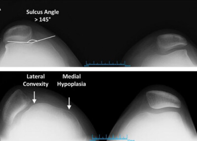
Image shows Sunrise radiographic views showing the Dejour classification system for trochlear dysplasia: (A) dysplasia type A with a shallow sulcus angle (right knee); (B) dysplasia type C with lateral convexity and medial hypoplasia of the trochlea (left knee).

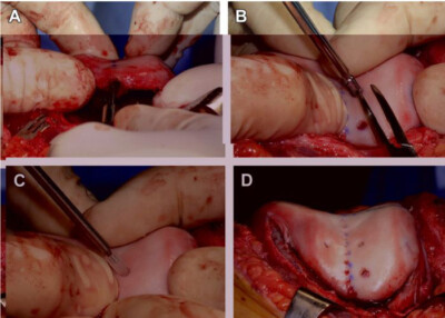
(A) The trochleoplasty is performed using a bur to facilitate creation of a decreased sulcus angle; a tap (B) is used to prepare attachment sites for bioabsorbable screws (C), which secure the deepened trochlea as it heals; (D) the completed sulcus-deepening trochleoplasty (left knee).
Are you a candidate for a trochleoplasty procedure?
There are two ways to initiate a consultation with Dr. LaPrade:
You can provide current X-rays and/or MRIs for a clinical case review with Dr. LaPrade.
You can schedule an office consultation with Dr. LaPrade.
(Please keep reading below for more information on this treatment.)
Trochleoplasty Procedure
A trochleoplasty is performed by reshaping the distal aspect of the femur. In this circumstance, guide pins are placed along the undersurface of the trochlea cartilage surface and a saw is used to undermine the articular cartilage. A V-shaped groove is then prepared in the distal femur, and the articular cartilage is positioned down into the V to reconstitute the bony groove. A trochleoplasty is rarely performed as a primary procedure, and most often is performed after patients have failed other types of reconstructions, including a MPFL reconstruction. Because of the complex nature of these problems, a trochleoplasty can also be commonly performed with a distal femoral osteotomy, a medial patellofemoral ligament reconstruction and/or a tibial tubercle osteotomy.
For this reason, the patient needs a thorough clinical exam, radiographic work up, and evaluation of the status of the articular cartilage prior to undergoing a trochleoplasty to obtain the best chance of an optimal outcome.
Dr. LaPrade published a study about Trochleoplasty and it can be found HERE
