Sports medicine knee specialist Robert LaPrade, MD PhD reviews how to read an MRI of a radial meniscus tear. It is important to be able to differentiate a radial meniscus tear on magnetic imaging because they are a lot different than other tears. In a younger patient a radial tear can be catastrophic because it completely destabilizes the meniscus.
To begin, Dr. LaPrade uses a coronal view of a right knee. In this specific case there is a radial tear of the anterolateral aspect of the lateral meniscus, which is a common location for these injuries. As the imaging moves deeper you begin to see signal intensity in the alterolateral of the lateral meniscus, where the tear is located. It is a subtle finding because this injury is not seen all well in the coronal view. You want to make sure there is not any extrusion of the meniscus and to make sure that the root attachment is still attached.
The next perspective is a sagittal view. As you move forward from the lateral side you start to see some appearance of the lateral meniscus and there is evidence for some disruption.
The best view to determine radial meniscus tears is the axial view. As you reach the center of the joint you can see a cut right through the center of the joint. The evidence of fluid indicates there is a complete disruption, which is a radial tear of the anterior aspect of the lateral meniscus. This tear makes the whole meniscus unstable and it does not act as a normal shock absorber.
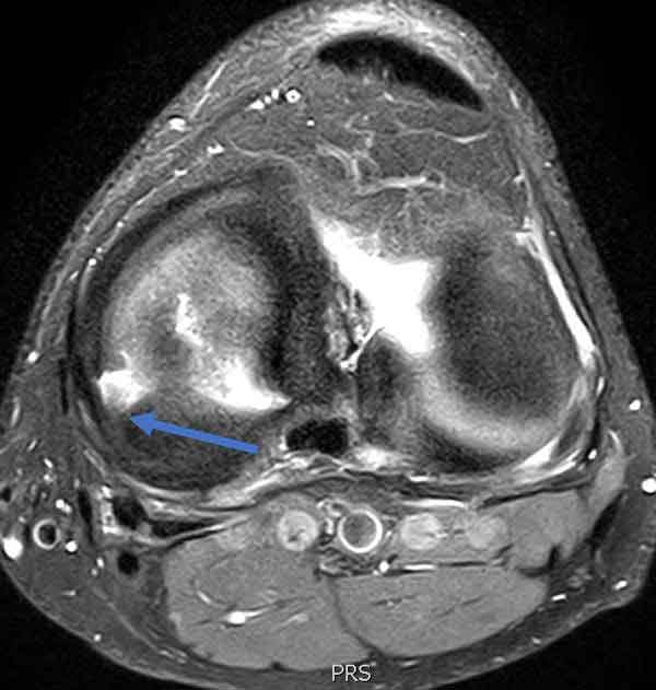
Lateral Meniscus Radial Tear
Axial view MRI scan demonstrating a complete radial tear of the lateral meniscus. The meniscus is essentially sliced in two and the edges tend to pull apart. These tears are particularly ominous. If the patient is young and or if there is fairly normal cartilage present, lateral meniscus radial tears should have an attempt at a repair. The success rate in a systematic review was approximately 80%, indicating that the surgery should be attempted to be performed in these patients (if possible).
CLICK IMAGE TO ENLARGE
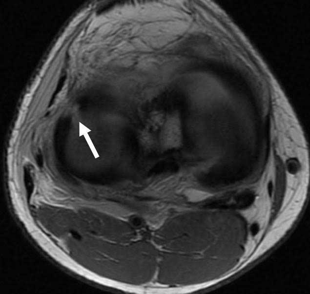
Radial Tear of Medial Meniscus
Axial MRI scan demonstrating a complete radial tear of the medial meniscus. The edges of the medial meniscus are separated, which effectively makes the medial meniscus nonfunctional. Similar to radial tears in the lateral meniscus, it is recommended that radial tears of the medial meniscus be repaired in patients that are young or even in older patients that have normal articular cartilage.
CLICK IMAGE TO ENLARGE