Description of Posterior Horn Lateral Meniscus Tear
The posterior horn of the lateral meniscus includes the main body of the lateral meniscus, posterior to the popliteus tendon, and its root attachment on the posterior aspect of the tibia.
The lateral meniscus is even more important than the medial meniscus for shock absorption. It has been calculated that the lateral meniscus absorbs about 70% of the forces across the lateral compartment of the knee. Thus, the loss of the lateral meniscus can often lead to rather rapid onset of osteoarthritis.
Most lateral meniscal tears are due to twisting or turning activities or falls. Lateral meniscal tears are common in sports such as skiing.
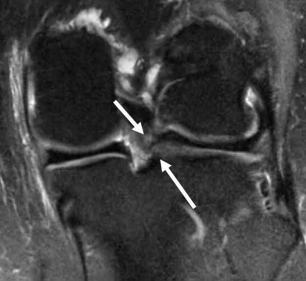
Coronal MRI scan demonstrating a posterior horn lateral meniscus type II root tear. The lateral meniscus root is torn up to 12% of the time concurrently with an ACL tear. Failure to recognize this tear pattern can lead to failure of the ACL reconstruction graft due to increase in stress with both anterior translation of the knee and for internal rotation during the pivot shift.
CLICK IMAGE TO ENLARGE
Symptoms of a posterior horn lateral meniscus tear:
- Pain
- Swelling and stiffness, increases gradually from hours to days after injury
- Catching or locking
- Instability
- An inability to straighten the knee
Have you sustained an injury to the posterior horn of the lateral meniscus?
There are two ways to initiate a consultation with Dr. LaPrade:
You can provide current X-rays and/or MRIs for a clinical case review with Dr. LaPrade.
You can schedule an office consultation with Dr. LaPrade.
(Please keep reading below for more information on this condition.)
Treatment for a Posterior Horn Lateral Meniscus Tear
In most circumstances, we strive to repair the posterior horn of the lateral meniscus, either with sutures, or with a pull through suture if it is a meniscal root detachment. In those circumstances where the tear is not repairable, which is more common than not, the patient should be followed very closely to determine if they are developing arthritis.
If the meniscus cannot be repaired, future treatment options include observation, bracing, activity modification, injections, or consideration for a lateral meniscal transplantation. A complete workup as to the patient’s suitability for any of these treatments necessary through a complete physical exam and any necessary radiographic (x-rays, MRI, etc.) work-ups.
Post-Op
We commonly counsel patients to let us know if they are experiencing any pain or swelling with activities, because these are the signs of early onset arthritis. The patient should be followed up routinely with standard x-rays, a Rosenberg view, and an assessment of whether they are developing joint space narrowing or bone spur (osteophyte) formation.
Related Studies
- Popliteomeniscal Fascial Tears Causing Symptomatic Lateral Compartment Knee Pain
- Anatomic Analysis of the Posterior Root Attachments of the Menisci
- Posterior Root Avulsion Fracture of the Medial Meniscus
- Not Your Father’s (or Mother’s) Meniscus Surgery
- Anterior Intermeniscal Ligament of the Knee – An Anatomical Study
- Prospective Outcomes Study of Meniscal Allograft Transplantation