Description of a Lateral Meniscus
The lateral meniscus is an essential shock absorber on the outside (lateral) aspect of the knee joint. It absorbs about 70% of the shock of the lateral compartment. Lateral meniscal tears are not as common as medial meniscus tears. This is because the lateral meniscus is more mobile and not secured as much to the lateral tibial plateau as the medial meniscus is to the medial tibial plateau. Thus, when there is a lateral knee injury such as a lateral meniscus tear, it is very important to try to repair the tear, because if not repaired and is trimmed out there will be an increase to the load on the lateral compartment, which ultimately leads to osteoarthritis.
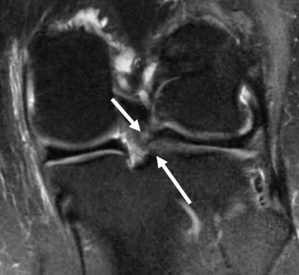
Coronal MRI scan demonstrating a posterior horn lateral meniscus type II root tear. The lateral meniscus root is torn up to 12% of the time concurrently with an ACL tear. Failure to recognize this tear pattern can lead to failure of the ACL reconstruction graft due to increase in stress with both anterior translation of the knee and for internal rotation during the pivot shift.
CLICK IMAGE TO ENLARGE
Symptoms of a lateral meniscus tear:
- Pain
- Swelling and stiffness, increases gradually from hours to days after injury
- Catching or locking
- Instability
- An inability to straighten the knee

X-rays show the results of removing a lateral meniscus in a very young patient with subsequent bone-on-bone arthritis of the outside (lateral) knee compartment on a left knee flexion view, providing objective information on the importance of repairing lateral meniscus tears whenever possible.
Treatment of a Lateral Meniscus Tear
Due to the added shock absorbing capacity of the lateral meniscus, it is essential to attempt to repair lateral meniscus tears if possible. We believe that trying to stimulate an improved healing environment through the use of bone marrow elements, platelet rich plasma (PRP), and a large number inside-out meniscal repair sutures, can lead to improved ability to heal these tears, especially in younger patients.
Post-Op
The treatment for patients who undergo a partial lateral meniscectomy is to initiate physical therapy on the first day after surgery. A treatment regimen working on reactivation of the quadriceps muscles, regaining of full knee and patellar mobility, and a quick resolution of knee swelling is emphasized. In general, we recommend that patients who have a minimal amount of lateral meniscus trimmed out hold back on any impact activities until a minimum of 6 weeks after surgery. In patients who have a significant amount of meniscus resected, it is often recommended to avoid significant impact activities due to the higher risk of the development of osteoarthritis in these patients with this activity.
When a torn lateral meniscus needs to be resected, we strongly recommend that these patients be followed very closely. Patients need to report back to their physician if they have any pain or swelling with activities, because these are the signs of arthritis and may indicate further progression of arthritic changes. If this is present, further treatment to include activity modification, low impact exercising, unloader braces, injections, or possible meniscal transplantation may be indicated.
It is almost inevitable that when one has a significant amount of the lateral meniscus resected that they will develop further arthritic changes over time. Because not everybody is the same, these changes can develop within a few weeks up to over a decade. We have observed that this often can develop rapidly in younger patients. Thus, one of the important things is to recognize that if one has any pain or swelling, they should follow up to make sure they are not developing any joint space narrowing or bone spurs, which would indicate that the lateral compartment articular cartilage is wearing out.
Lateral Meniscus Tears FAQ
The lateral meniscus is almost a C-shaped cushion on the outside of one’s knee. The lateral meniscus absorbs up to 70% of the shock, on average, that is placed across the lateral compartment of the knee. Therefore, the lateral meniscus is very important to preserve when it is torn because there is a very high risk of progression of arthritis if a meniscus is not repaired.
1. Can a lateral meniscus tear heal itself?
There are some types of lateral meniscus tears that could potentially heal themselves. They depend upon the type of meniscus tear and the location of the tear in the joint. If the meniscus is torn at the edge where it attaches to the joint lining and the tear is small, there is the possibility this could heal. In addition, there have been occasional case reports that report that some radial tears may heal over time, although this is not expected for the vast majority of tears. What it does indicate is that the lateral meniscus does have some ability to heal, so pushing the limits for a repair, especially in a young patient, would be indicated.
2. Where does the knee hurt when there is a lateral meniscus tear?
In general, a lateral meniscus tear should cause pain along the joint line of the outside part of the knee. In addition, one could have pain in the back of their knee with deep squats or they could have pain on the outside of their knee when they put their leg in a figure-of-4 position. These are the usual locations that a lateral meniscus tear will hurt.
3. Is the lateral meniscus tear worse than a medial meniscus tear?
It is hard to differentiate what type of tear is worse if it is repairable. However, it is well known that if a lateral meniscus is taken out, the consequences are almost always worse than having a medial meniscus resected.
4. What does a complex lateral meniscus tear mean?
A complex lateral meniscus tear can include a radial tear, a radial flap tear, a tear that is chewed up and macerated, or a root tear. In general, a simpler type tear would be one that is very small and trimmable or one that is torn at the meniscus attachment to the joint lining, which is easily repairable.
5. How does one treat a lateral meniscus tear?
In general, because of the important shock-absorbing ability of the lateral meniscus, repairable lateral meniscus tears should have an attempt at a repair as long as the patient is fairly active and does not have any significant arthritis. For smaller tears or tears that are in an area with hardly any blood supply, a trimming would possibly be indicated. This would be called a partial lateral meniscectomy.
6. What type of cysts are those that develop with a lateral meniscus tear?
Sometimes a lateral meniscus will have a split in the middle, called a horizontal tear, whereby fluid can leak out through the joint lining. This fluid can accumulate over time and almost be a jelly-like substance. This is called a lateral meniscal cyst or a parameniscal cyst. In most circumstances, the cyst is caused by the meniscus tear. Therefore, when one does excise the cyst, the meniscus tear needs to be addressed, most commonly by suturing the meniscus tear to prevent the cyst from reforming.
7. What does one do tor the lateral meniscus tear when one has osteoarthritis?
The osteoarthritis amount would determine what type of treatment may be indicated for a lateral meniscus tear. If there is very minimal arthritis and the meniscus is repairable, a repair should be performed. However, if there is bone-on-bone arthritis and there is a tear present, unless there is a definitive mechanical problem, limiting one’s function, and observation, rather than a partial meniscectomy may be indicated.
8. How does one treat an anterior horn lateral meniscus tear?
Anterior horn lateral meniscus tears are harder to treat technically because the surgical instruments that have been developed are not as adept at addressing these tears. For trimmable tears, using a curved shaver or an arthroscopic device that bites backward, called a backbiter, may be indicated. If there is a tear of the meniscus that is repairable, using a technique called the outside-in technique would be advisable to perform a direct repair of the anterior horn lateral meniscus tear.
9. How often are lateral meniscus tears missed on MRI?
In our practice we find that MRI is much more successful at diagnosing medial meniscus tears compared to lateral meniscus tears. In the literature, the lateral meniscus tear diagnosis via MRI has been reported to be between 80% to 90% successful. Thus, it is entirely possible that one could go into surgery thinking that the lateral meniscus is okay and a further evaluation and probing may reveal a large tear that was not picked up on the MRI scan.
10. What does one do to a lateral meniscus tear with extrusion?
Extrusion can be due to 2 things. One could be that the cartilage is worn out and the meniscus is being squirted out of the joint because of the amount of arthritis. The second could be that there is a radial root tear of the lateral meniscus which is causing it to slip out of the joint. Radial or lateral meniscus root tears should be repaired if the cartilage surfaces are still pretty good. This involves releasing scar tissue and pulling the meniscus back in the joint and then either suturing it together or tacking it back down to bone, depending upon the type of tear.
11. What is a lateral meniscus tear in the red zone?
Meniscus tears are classified according to the distance from the joint lining, which is almost directly proportional to the blood supply that is present. Within the first 2-3 mm there is a good blood supply, so tears in this zone we call red-red. Tears that are 2-3 mm away from this are more at the edge of the red zone and into the white zone where there is less blood supply and we will call these red-white tears. Tears that are further along, more than 6 mm from the edge, are more at an area where there is poor blood supply and these are called white-white zone tears. More recent literature suggests that the meniscus does have a good ability to heal itself with white-white zone tears, so repairs of large tears in this zone would be recommended.
12. How does one differentiate between a lateral meniscus tear and iliotibial band friction syndrome?
One of the best ways to differentiate between a lateral meniscus tear and iliotibial band friction syndrome is performing a good history and physical examination on a patient. In general, iliotibial band friction syndrome develops over time with activity, usually after a runner has been running up to 2 miles and does not hurt initially. Meniscus tears that are catching the joint would be expected to hurt as soon as one started to run. In addition, meniscus tears should be painful at the joint line, whereas a tight iliotibial band would usually hurt when one pushes directly over the lateral epicondyle and flexes and extends the knee. The iliotibial band that may be irritated with the iliotibial band friction syndrome should be painful with this maneuver if one pushes down hard enough on the lateral epicondyle.
13. Can a lateral meniscus tear cause a Baker’s cyst?
A Baker’s cyst is a fluid pocket that develops when fluid leaks out the back of the inside part of the knee between the medial head of the gastrocnemius and the direct arm of the semimembranosus. Most of us have a little hole in the back of our knees at dislocation. Thus, anything that could cause swelling in the knee can result in a Baker’s cyst forming. Thus, if a lateral meniscus does cause some swelling, it can lead to the development of a Baker’s cyst. The treatment for a Baker’s cyst is to treat the problem at the front of the knee and other than very rare occasions, surgery to treat a Baker’s cyst is not performed.
14. Can stem cell treatment heal a lateral meniscus tear?
There are many different types of lateral meniscus tears with the importance depending on the size of the tear, the location in terms of blood supply, and its duration. For large flaps of meniscus tears or meniscus root tears which have separated, a bone marrow aspirate injection or a true stem cell injection, which are not allowed in the U.S. and mainly performed in Europe or Chile, would have a low chance of working. We do know that bone marrow injections can make a knee less irritated, but the chance of healing a tear in this circumstance would be minimal. For meniscus tears that do have a high potential of healing on their own, such as small tears at the meniscocapsular junction, it is theoretically possible that the bone marrow injections, which are commonly called “stem cell injections” in the United States, could potentially lead to their healing. However, larger tears would still be suited to having a repair and possible augmentation with bone marrow aspirate.
15. What is a lateral meniscus free-edge tear?
If one slices through a meniscus, it is shaped like a triangle. The inner portion of the triangle that is thinner is just like the inner portion of the meniscus. This would be the free-edge tear. At this location the meniscus may only be 1 mm to 2 mm in total depth, so an attempt at a repair would not be indicated. In most of these small free-edge tears, trimming them carefully to remove the area of the tear, but not remove any further tissue, would be indicated.
16. What should one do with a lateral meniscus tear in a teenager?
Because of the severe consequences of taking a lateral meniscus out in a teenager, which almost always leads to the development of osteoarthritis within 1-2 decades, it would be recommended to perform a repair if possible. Many complex tears may be suitable to attempted repairs at this age group because of their better healing potential and also may benefit from biologic augmentation with a marrow venting procedure, bone marrow injection, or a leukocyte-poor PRP injection to augment the healing process.
17. What causes locking with a lateral meniscus tear?
One of the most common causes of locking with a lateral meniscus tear are either a tear that has torn at the edge and slips into the joint with twisting and turning or a meniscus that totally flipped from the back to the front, called a bucket-handle tear, which can block extension. Many athletes that are seen that do present with a a bucket-handle tear had a previous history of catching or locking with activities that was a prelude to having the entire meniscus flip upon itself.
18. What can be done for a horizontal lateral meniscus tear?
Horizontal lateral meniscus tears can be repaired and this could be considered in younger patients. In older patients who have symptoms, removing the inferior leaf of the tear and preserving the superior leaf to try to serve as shock absorption would be recommended. We do know that removing half of the meniscus definitely increases the risk of arthritis, but it would be better than removing the whole meniscus. A horizontal meniscus repair would involve cleaning out between the two upper and lower portions of the meniscus with a shaver, suturing the top and the bottom together, and consideration of inserting a fibrin clot between the two, like a peanut butter and jelly sandwich, to try to deliver the maximum amount of growth factors into the area of the tear to try to maximize healing.
19. What should be the treatment for a lateral meniscus bucket-handle tear?
Because the lateral meniscus is so important for shock absorbing, one should attempt to have a repair of the lateral meniscus when possible and when one does not have any significant arthritis. This would involve an arthroscopy to push the meniscus back in place and using multiple sutures to hold the meniscus in place. The gold standard for this would be an inside-out repair with multiple sutures, varying anywhere from 10-20 total number of sutures.
20. What causes a lump along the joint line with a lateral meniscus tear?
The most common cause of a lump with a lateral meniscus is a lateral meniscus cyst at the joint line. Small lumps can occur from a fragment of the meniscus being displaced along the joint line or under the meniscus and along the capsule, which can sometimes be palpable and painful.
Have you sustained a lateral meniscus tear?
There are two ways to initiate a consultation with Dr. LaPrade:
You can provide current X-rays and/or MRIs for a clinical case review with Dr. LaPrade.
You can schedule an office consultation with Dr. LaPrade.