PCL Stress Radiographs
PCL stress radiographs are obtained to objectively determine the amount of increased posterior tibial translation in a patient that may have a possible PCL tear preoperatively and also to determine the postoperative surgical outcomes. PCL stress radiographs can objectively measure the amount of increased posterior tibial translation on the injured knee compared to the contralateral normal knee. This is important because it has been documented in the peer reviewed literature that the amount of posterior translation assessed on a clinical posterior drawer test is not always accurate.
The important landmarks that are utilized for PCL stress x-rays are
- 1) the top of Blumensaat’s line, which is a point at the top of intercondylar notch
- 2) perfectly overlapping medial and lateral femoral condyles, and
- 3) a tibia that is not rotated compared to the contralateral side. In addition, the knee should be flexed to 90 degrees over slightly more, but it should not be in a more extended position (like 80 degrees) because this would result in a false underestimation in the amount of posterior tibial translation that occurs.

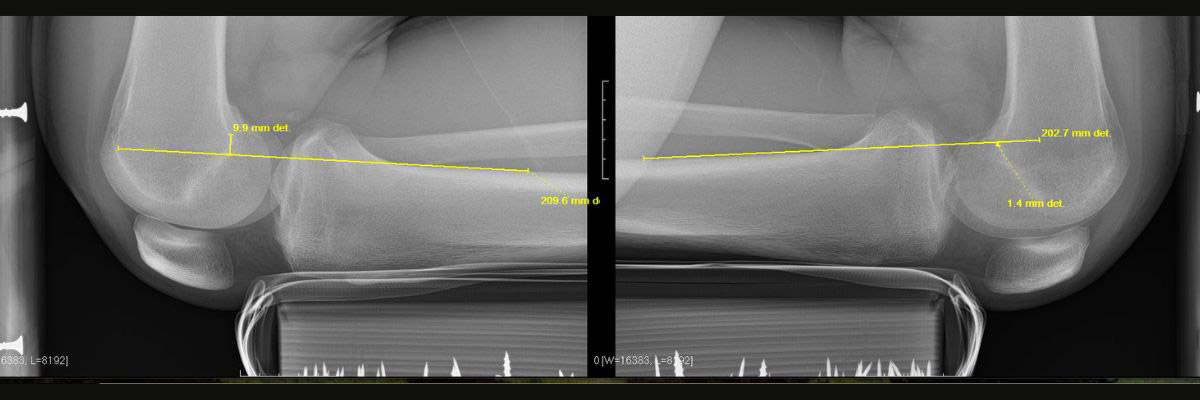
Figure 1. Bilateral PCL stress x-rays demonstrating 11.3 mm of increased posterior tibial translation on the injured compared to the normal contralateral knee. This is consistent with a high-grade PCL tear.
When the x-ray technique is optimized, a line is drawn along the posterior cortex of the tibia which goes through the femur. A perpendicular line is then drawn up or down to Blumensaat’s point. The measurements are then compared to the contralateral knee. Stress radiographs that are less than 4 mm are felt to be a very minor PCL or possibly normal laxity, while 8 mm or more is consistent with a complete nonfunctional PCL tear. In particular, stress radiographs that are greater than 12 mm of increased posterior tibial translation are consistent with either a combined PCL and posterolateral or posteromedial corner injury or in a patient with a very flat tibial slope.
It is important to recognize that the techniques of PCL stress radiographs can be difficult to obtain initially when working with one’s radiology technicians. The surgeon should meet with the technicians to review what the optimal landmarks are to ensure that objective information is obtained to best treat a patient’s pathology.
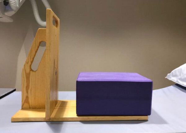
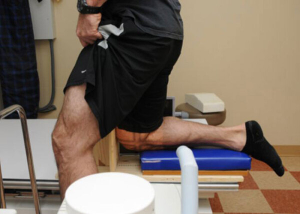
The patient should kneel on a foam bolster with their knee flexed to 90 degrees and the bolster should be positioned at the tibial tubercle. This will allow for the knee to have appropriate amounts of translation in a PCL injured knee. A PCL stress x-ray device can be fabricated to do this, but if the device is not available, a thickened piece of foam can also be utilized for the patient to kneel on to obtain the stress radiographs.


Figure 2: PCL Stress Device
Figure 3: Example of a PCL kneeling stress x-ray device with the patient kneeling on his right knee.
