Description of Genu Recurvatum
One of the more difficult problems to treat in sports medicine is genu recurvatum.
In this condition, an athlete sustains an injury and has excessive backwards motion of their knee (hyperextension of the knee). In these circumstances it can often be difficult to treat this problem and patients can have significant disability.
It is important to differentiate this problem from patients who may have had a growth plate injury when they were young and now have bony problems with recurvatum or in patients with polio or other muscle diseases where their quadriceps are so weak they have excessive hyperextension of the knee.
These injuries are most commonly caused by a blow to an extended knee with a subsequent injury to either some of the main knee structures or possibly just the structures of the posterior aspect of the knee.
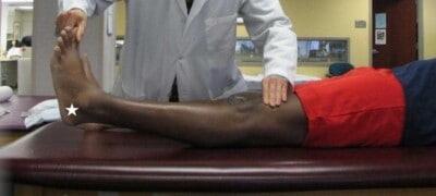
Clinical exam demonstrating a significant increase in recurvatum, or heel height, in a patient with a posterolateral corner injury of the knee. Increases in heel height associated with genu recurvatum are usually associated with a combined ACL and posterolateral corner injury, especially with either an isolated fibular collateral ligament tear or a fibular collateral ligament tear with a biceps femoris tendon avulsion off the fibular head.
CLICK IMAGE TO ENLARGE
Other causes of Genu Recurvatum Include:
- A defined disorder of the connective tissue
- Laxity of the knee ligaments
- Instability of the knee joint due to ligaments and joint capsule injuries
- Irregular alignment of the femur and tibia
- A deficit in the joints
- A discrepancy in lower limb length
- Certain diseases: Cerebral Palsy, Multiple Sclerosis, Muscular Dystrophy
- Birth defect/congenital defect
Symptoms of Genu Recurvatum:
- Knee giving way into hyperextension
- Difficulty with endurance activities
- Pinching in the front of the knee
A careful evaluation must be performed to determine if there is any cruciate ligament tear, posterolateral corner injury, or a medial knee injury to include the superficial medial collateral knee and posterior oblique ligament. In some circumstances, none of these more commonly known structures may be injured and all of the injured structures are in the back of the knee.
Our anatomic and biomechanical research has discovered a tibial attachment of the oblique popliteal ligament which, when injured, can result in increased knee hyperextension.
The best way to clinically diagnose the amount of hyperextension of the knee is to measure the patient’s heel heights. If there is a normal contralateral (opposite) knee to compare to, an increase in heel height can be diagnostic for genu recurvatum.
A careful assessment of a patient’s overall knee alignment must be performed via a long leg x-ray. Their posterior tibial slope must also be calculated on a lateral knee x-ray. Patients who have a decreased posterior tibial slope tend to have more problems with knee hyperextension than those who have increased posterior tibial slopes. We have also discovered this as part of our research.
Are you experiencing genu recurvatum symptoms?
There are two ways to initiate a consultation with Dr. LaPrade:
You can provide current X-rays and/or MRIs for a clinical case review with Dr. LaPrade.
You can schedule an office consultation with Dr. LaPrade.
(Please keep reading below for more information on this condition.)
Treatment for Genu Recurvatum
If a patient does not have an associated cruciate ligament and/or collateral knee injury present, the usual treatment is to attempt a rehabilitation program to see if the patient can improve their overall quadriceps strength to compensate for the symptomatic knee hyperextension. If this does not work, then possibly a biplanar proximal tibial osteotomy, where the patient’s posterior tibial slope is increased, may be indicated. While these are extensive surgeries, they have been well documented to decrease knee hyperextension and allow patients to return to a high functioning level.
Our treatment for isolated genu recurvatum is a proximal tibial anteromedial or anterolateral osteotomy that increases the patient’s posterior tibial slope. These surgeries have been found to be very effective in decreasing a patient’s knee hyperextension and returning them to increased activities after the osteotomy heals.
Post-Operative Protocol for Genu Recurvatum
Something that is most important in a patient who presents with genu recurvatum is to combine the clinical exam with above noted radiographs to devise a treatment plan. If a patient does not have any obvious collateral or cruciate ligament injury, then a closely supervised quadriceps strengthening physical therapy program may be indicated. For those patients who have already enrolled in a rehabilitation program and/or have continued problems with knee hyperextension, a brace that attempts to prevent hyperextension of the knee may be indicated. In our hands, we have found that almost all patients who have this problem may respond well for a brief period of time to a brace that limits hyperextension, but in general, still have problems and need to consider surgery.
Genu Recurvatum FAQ
1. What is genu recurvatum?
Genu recurvatum is a term that is used when one hyperextends their knee. Knee hyperextension can be caused by several causes. These include muscle weakness, especially of the muscles in the top of the thigh (quadriceps), it can be due to injury, or it can occur due to the shape of one’s bones at their knee.
2. I have genu recurvatum. How to fix genu recurvatum?
The most important thing about treating genu recurvatum is to try to determine the cause of it. If it is due to a muscle weakness issue, then a program of muscle strengthening or bracing may be indicated. In the past, genu recurvatum was very common because polio caused weakness of the quadriceps muscles. If it is due to an injury, and one finds they have genu recurvatum right after an injury, it is important to determine the specific structures that were injured and whether they can be repaired or reconstructed to address the knee hyperextension. We have found that patients with a combined ACL tear and an injury to the lateral (fibular) collateral ligament have about 3 cm of increased heel height. Treating this type of genu recurvatum is relatively easy and consists of reconstructing both the ACL and the LCL. If one has a chronic injury, or has a bony problem with the tibia, the only solution may be bracing or a surgery where the sideways angle of the tibia, its sagittal slope, is increased with an osteotomy (where one breaks a bone and changes the angle of the shin bone).
3. How successful is genu recurvatum surgery?
The most important thing about genu recurvatum surgery is to make sure that the clinical diagnosis is correct. Most commonly, patients present with a chronic injury who have their knee bend backwards have functional issues which can be disabling. In most of these patients, because the injury is chronic, a workup will determine that their sagittal plane tibial slope is too flat and an osteotomy is indicated. We have found that a proximal tibial osteotomy which slopes the tibia posteriorly is very effective to address these cases of symptomatic genu recurvatum.
4. How common is genu recurvatum and who treats these?
Genu recurvatum cases are uncommon and because of this they are often not diagnosed correctly. For this reason, most cases are treated at referral centers. In addition, because they are uncommon, many physicians may feel uncomfortable at performing an osteotomy to address the recurvatum. That is one of the more common issues that we see in patients that are referred to us. However, in effect, the main treatments for genu recurvatum are strengthening the quadriceps muscles, using a brace to prevent hyperextension (which is often quite bulky and not tolerated well by patients) or proceeding with a proximal tibial osteotomy which increases the slope of the tibia to decrease or eliminate the genu recurvatum.
5. How can one determine the severity of their genu recurvatum?
Answer: The best way to determine the extent of one’s genu recurvatum is simple. Obtaining a ruler and measuring one’s heel height, where the thigh is held against the examining table and one’s great toe is lifted up, and measuring the distance in cm between the bottom of the heel and the table, it is the most effective way to determine the heel height, which correlates to the amount of genu recurvatum. In general, 1 cm heel height correlates to 1 degree of hyperextension, which is denoted by a negative sign for knee motion examination purposes. Therefore, somebody who has “-5” degrees hyperextension would have a 5 cm heel height.

