What is an MCL Tear?
The most common knee ligament injury is an injury to the medial aspect of the knee. There are three main anatomic structures on the medial side of the knee, with the medial collateral ligament (MCL) being the largest and strongest. A large number of isolated MCL medial knee injuries are due to sporting events. They can be either a contact or non-contact stress to the outside of their knee, which in turn stretches or tears the medial knee structures on the inside of the knee.
The grade of the medial knee injury is based upon the amount of tearing present and treatment options depend on the location of the tear and if other ligaments are concurrently injured.
- Grade I: partial MCL tear
- Grade II: near complete MCL tear
- Grade III: a complete MCL tear – the ligament is non-functional
Dr. LaPrade will perform a MCL reconstruction on patients who exhibit a grade III tear. In addition, it is well recognized having any “looseness” of the medial knee structures can cause an ACL graft to fail. Thus, when there is a combined medial knee injury and ACL injury, it is important to make sure the MCL injury heals completely prior to the ACL reconstruction or it should be concurrently repaired or reconstructed.
In the case of very severe combined knee ligament injuries, especially with a PCL tear, a concurrent medial knee injury should undergo a repair, augmentation repair, or a complete medial knee reconstruction (MCL reconstruction surgery).
What is MCL Surgery?
Dr. LaPrade has performed extensive anatomic, biomechanical, diagnostic and other related studies to better understand medial knee injuries and structures. Through this work, he has been able to develop an anatomic medial knee reconstruction procedure that has been performed in patients and is currently undergoing clinical outcome studies.
Historically, MCL reconstruction surgery of medial knee injuries resulted in a significant amount of patients developing postoperative stiffness that often results in more surgeries. Thus, we have developed newer techniques which allow us to have patients move their knee sooner to try and decrease the risk of stiffness and the necessity of secondary surgeries. For all medial knee injuries, a careful assessment must be performed with a thorough physical examination, the use of stress x-rays and the use of high field strength MRI to identify any concurrent injuries.
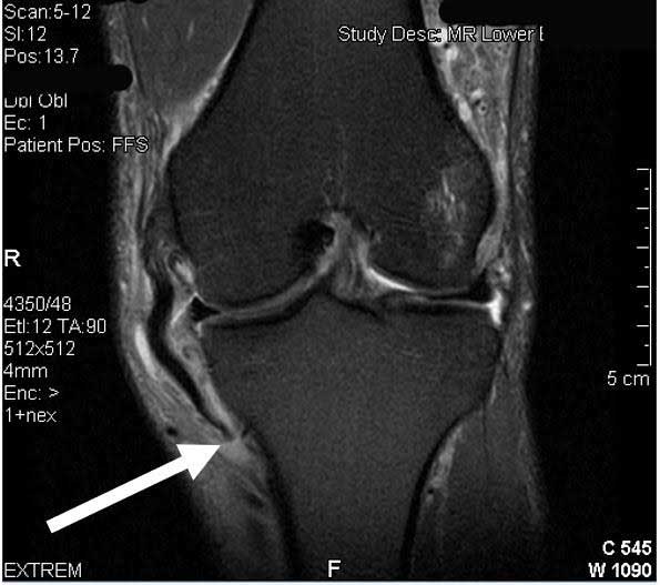
Coronal MRI scan demonstrating an MCL tear off the tibia (meniscotibial based). Meniscotibial based MCL tears have a lower chance of healing, especially when the knee has a significant increase in external rotation or when it gaps in full extension. In some of the semeniscotibial-based MCL tears, the torn MCL flips over the pes anserine hamstring tendons and have no chance of healing back to the tibia In these circumstances, there is a very high chance that the MCL will not heal and will require surgery due to continuing instability.
CLICK IMAGE TO ENLARGE
Are you a candidate for MCL Reconstruction?
There are two ways to initiate a consultation with Dr. LaPrade:
You can provide current X-rays and/or MRIs for a clinical case review with Dr. LaPrade.
You can schedule an office consultation with Dr. LaPrade.
(Please keep reading below for more information on this treatment.)
How Long is Recovery Time After MCL Surgery?
In the acute situation, Dr. LaPrade generally places patients into an early rehabilitation program to emphasize quadriceps reactivation, edema control and knee range of motion. For isolated acute MCL ligament injuries, most athletes can return to sports by multiplying the grade of the injury by two (in weeks) as a general time frame. Thus, a grade I acute MCL injury usually needs 1-2 weeks to heal, while a grade II injury takes 3-4 weeks to heal and a grade III isolated complete MCL ligament injury typically takes 5-6 weeks of properly guided rehabilitation to have the injury heal completely.
The use of an MCL protective knee brace is also commonly recommended in the acute situation for medial knee injuries when the knee is significantly unstable. Thus, we recommend the patient be fitted by a brace specialist who understands the patient’s goals and will properly fit the MCL brace which is durable enough for their desired activity levels.
Related Studies
- sMCL – Anatomic Augmented Repair vs Anatomic Reconstuction
- Development of an Anatomic Medial Knee Reconstruction
- Structural Properties of Primary Medial Knee Ligament
- Anatomy of the Medial Part of the Knee
- Correlation of Valgus Stress Radiographs
- Medial Knee Part I – Static Function of the Main Medial Knee Structures
- Management of Injuries to the Medial Side of the Knee
- Analysis of Anatomical Medial Knee Reconstructions
MCL Injury FAQ
1. Can the MCL heal on its own?
The MCL is surrounded by other tissues and does have a good ability to heal on its own when it is torn. However, there are some types of MCL tears that commonly do not heal well and need to be followed more closely. These include MCL tears with a multiligament knee injury, MCL tears that are completely torn off of the femur whereby the femur gaps to the outside when the knee is out straight, and those that tear off the tibia. These 3 MCL tears in particular have a much lower chance at healing and need to be followed very closely to see if they do heal. Sometimes proceeding directly to surgery, with the amount of instability that a patient presents with or with a particular type of tear pattern which shows up on the MRI, may be indicated for these circumstances.
Coronal MRI scan demonstrating an MCL tear off the tibia (meniscotibial based). Meniscotibial based MCL tears have a lower chance of healing, especially when the knee has a significant increase in external rotation or when it gaps in full extension. In some of the semeniscotibial-based MCL tears, the torn MCL flips over the pes anserine hamstring tendons and have no chance of healing back to the tibia In these circumstances, there is a very high chance that the MCL will not heal and will require surgery due to continuing instability.
CLICK IMAGE TO ENLARGE
2. Can an MCL tear result in one being able to not bend or straighten their knee?
MCL tears are a particular type of knee ligament injury whereby one can lose motion after an injury. Therefore, it is entirely possible that as one heals that scar tissue may form and one may either not be able to bend or straighten their leg. This is why it can be particularly important to make sure one is in a physical therapy regimen when one does tear their MCL to maximize the chance that it will both heal and also heal without having a stiff knee
3. How long does it take an MCL tear to heal?
The time that it takes an MCL to heal usually depends upon the amount or grade of the tear. A grade I mild intrasubstance tear with no gapping in the knee usually takes 1 to 2 weeks to heal in athletes. A partial tear, which we call a grade 2 tear, whereby there is some gapping present due to the tear towards the outside of the knee, but it is not completely torn, usually take 3 to 4 weeks. A complete tear, when it does heal, usually takes 5 to 7 weeks to heal in an athlete. If one is not wearing a brace or does not have a physical therapy regimen that addresses an MCL tear, then one may take longer to heal. In particular, tears that completely tear off the femur, whereby the knee gaps open with the knee out straight or ones that tear off the tibia and retract towards the knee joint, have a much higher chance of not healing.
4. Where is an MCL located?
The MCL is located on the inside of the knee. It is a thick ligament that is approximately 10 to 12 cm long. It prevents the knee from gapping towards the outside.
5. Where does MCL pain occur?
Because the MCL is on the inside of the knee, it will usually be on the inside of the knee where the pain is located. MCL tears can occur at their attachment on the femur (thigh bone), midsubstance, or towards the tibia. One’s clinician should be able to palpate after a tear and determine the location of the tear. If indicated, an MRI scan can correlate also with the location of one’s pain.
6. What are the symptoms of an MCL tear?
The symptoms of an MCL tear include pain on the inside of the knee, often in a rectangular shaped area, right on the direct inside or medial part of the knee, and a feeling of instability or true instability towards the outside of one’s knee. In addition, because the MCL can tear and put extra stress on the outside of the knee, some patients will have pain on the outside of the knee, either due to a bone bruise or a lateral meniscus tear. This should be evaluated closely by one’s physician.
7. How does one quantify an MCL tear?
The best way to quantify an MCL tear objectively is to obtain stress x-rays. Grade 1 MCL tears should have no gapping. Grade 2 MCL tears should have 1 to 2 mm of gapping from the injury compared to the contralateral knee, whereas a complete MCL tear should have 3.2 mm or more of gapping of the medial compartment on the injured knee compared to the contralateral knee. The old AMA Guidelines from 1966, which described 0 to 5 mm, 5 to 10 mm, and greater than 1 cm of gapping for grades 1, 2, and 3 MCL tears are useful only for subjective grading of an MCL tear and are not useful for the true objective amount of gapping that may occur in a patient.
8. Why does my MCL still hurt?
Some patients can have an MCL hurt for a period of time after the MCL is “healed” and do not have any increase in knee gapping. This is because of scar tissue that can occur, the potential of a deep MCL tear which can still cause some pain, and the possibility that bone may have formed instead of scar tissue within the MCL itself. This can also lead to pain. This is called Pellegrini-Stieda calcification and commonly occurs in grade 3 MCL tears.
9. When is the time to see a doctor with an MCL tear?
If one feels that their knee is unstable, especially with side-to-side gapping, one should see a physician to check on their MCL. This is to ensure that they receive proper treatment and therapy for the MCL tear, but also to make sure that there aren’t other injuries that occurred with the MCL tear which could be problematic if not treated further down the line.
10. When is the MCL the tightest?
The MCL is the tightest at 20 degrees of knee flexion. This is where it is the main structure that prevents one’s knee from gapping towards the outside.
11. When should one start rehabilitation for MCL tears?
One should start rehabilitation for MCL tears as soon as the injury is identified. One could work on motion, decreased swelling, and making sure that one’s muscles start to recover immediately. Complete MCL tears should probably always be treated with a brace to prevent them from healing in a looser position, whereas physical therapy for most MCL tears can be started immediately after the injury is diagnosed. This can include the use of a stationary bike, which we have found in athletes is the best way to get these to heal, as well as other exercises.
12. What happens when an MCL has a pop noise?
If one has an MCL tear and they hear a pop, the pop may be a bone bruise from the MCL gapping on the inside and compressing the bone on the outside of the knee. These lateral compartment bone bruises can result in one feeling a pop deep within their knee.
13. Which is worse–an ACL or MCL tear?
In general, we would say that an ACL tear is worse because most ACL tears do not heal and lead to instability, which requires surgery. Contrarily, MCL tears usually heal, but not always and a proper rehabilitation program can be useful for this.
14. Which is worse? An LCL tear or an MCL tear?
A complete tear of the LCL has a much lower chance of healing with the amount of healing in the literature being approximately 10% to 15%. MCL tears usually heal, probably 90% to 95% of the time, so if one does have an LCL tear, there is a much higher risk that they will have the LCL heal in a loose position and require surgery.
15. How do MCL injuries occur?
Most MCL injuries occur due to one stressing the inside of the knee. This can include a contact injury on the outside of the knee while they are playing sports or stepping in a hole and falling down towards the inside of their knee.
16. What is an MCL sprain?
In laymen’s terms, a sprain is equivalent to a tear. Thus, a sprain is basically talking about the same information as a tear. A grade 1 sprain would be a partial tear that does not gap open; a grade 2 sprain would be a partial tear that does have some instability; while a grade 3 sprain would be one where the MCL is completely torn and the knee gaps open to the outside.
17. What does the MCL stand for?
The MCL stands for the medial collateral ligament.
18. What type of surgery is performed for an MCL tear?
In our hands, we have developed an anatomic-based reconstruction of the MCL for when it tears. In the peer-reviewed literature, it has been found that MCL reconstructions do much better than repairs, so in most circumstances we recommend a complete reconstruction of the MCL to give the patient the best chance of having stability of their knee over time. This includes placing a graft at where the native MCL was located, called an anatomic reconstruction, and then pairing this with an early range of motion rehabilitation program to ensure that one does not have a stiff knee and has the best outcome over time.
19. Are MCL and meniscus tears the same thing?
The MCL is the main ligament on the inside of the knee which prevents the knee from gapping open. The medial meniscus is the cushion on the inside of the knee. While the pain can often be at the joint line for either of these, medial meniscal tears are not common with most MCL injuries and may be present only 10% to 15% of the time.
20. What does an MCL look like on an MRI scan?
The medial collateral ligament is located on the inside of the knee. On MRI scans, the most useful scan to look at is the coronal scan to look at the MCL along its course. The MCL attaches to the femur just behind the medial epicondyle and then courses down towards the tibia. On the tibia, it has 2 attachment sites. The first attachment site is over soft tissue, most commonly the ending of the anterior arm of the semimembranosus tendon, about 15 to 18 mm distal to the joint line. The main substance of the MCL, and also its strongest attachment point, is located 6 cm distal to the joint line. MRI scans are very useful to delineate the normal anatomy of the MCL and also to help to determine if it is torn off the femur, midsubstance, or off the tibia (or any combination of these).
21. Can one have an MCL tear without pain?
It is unlikely that one would have a new MCL tear without pain. This is because when one tears their MCL, even for a grade 1 tear, there is some intrasubstance tearing of the ligament which would cause some localized damage to the collagen fibers and usually result in some pain and swelling at that location. However, if one possibly had an MCL tear in the past and reinjured themselves, they may not have pain the second time around as much as they would have had with the first injury.

MCL tear off tibia (arrow) left knee
22. What is the treatment for MCL tear with an ACL tear?
The treatment of the combined ACL and MCL tear usually depends upon the physical exam. If both are torn and the knee does not gap to the outside, called valgus gapping in full extension, there is usually a good chance that the MCL will heal with 4 to 6 weeks of rehabilitation and then the ACL can be reconstructed after this. However, if the MCL is torn off the tibia, or if the MCL is completely torn at the femur and the knee gaps in full extension, then there is a much lesser chance that the MCL will heal. While trialing a rehabilitation program may be useful to help the patient get their range of motion back and get the swelling down, these patients have to be followed much more closely and evaluated for a combined ACL and MCL reconstruction.
23. When should an MCL be repaired with an internal brace?
There is not a lot of science to undergoing an MCL repair with an internal brace or a suture tape. The use of synthetic ligaments in the orthopaedic literature has almost always done poorly when they are followed over time. Therefore, our recommendation would be to consider surgery to reconstruct the torn MCL rather than doing a repair with an internal brace.
24. What happens when there is an MCL tear with an avulsion fracture?
Avulsion fractures of the MCL off the femur can occur, especially in younger patients. One needs to evaluate the amount of bone that is torn off with the avulsion fracture, as well as the amount of gapping that may be present on the physical exam, to determine if a fixation of the fracture fragment may be indicated to stabilize the MCL. In addition, one should also use the MRI to determine if there is an intrasubstance injury to the MCL with the fracture or if the MCL tissue looks good, an avulsion fracture fixation may be indicated.
25. What types of braces are best for MCL tears?
We believe that complete tears of the MCL should be braced such that one does not have the MCL heal with some looseness, which could give one symptoms over time. Providing a hinged knee brace, which often should be best fit to a patient’s knee, provides the best chance of providing stability to the knee such that one does not have the MCL heal loose, which could lead to further problems down the line.
26. What is the origin and insertion of the MCL?
The attachment site of the MCL on the femur is in a little bony depression or saddle, located behind the medial epicondyle. The attachment sites in the tibia are located about 15 to 18 mm distal to the joint line, over soft tissues, which commonly include the anterior arm of the semimembranosus, and then a very thick and strong attachment to the tibia further down from the joint line, with the main substance being 6 cm distal to the joint line.
27. How does the MCL tear while skiing?
An MCL is one of the more common injuries that occurs with skiing. This is because one can fall towards the inside of their knee, often having the skis not give way, and cause extra stress on the inside of the knee, which can tear the MCL. Many of these MCL tears while skiing do have other associated injuries, so an examination by a physician may be indicated if one feels that their knee is unstable.
28. Is it the MCL or TCL?
It is common in sports medicine to call all the structures on the inside of the knee the MCL. The main structures on the inside of the knee are the superficial medial collateral ligament, which in the lay press is called the MCL, as well as the deep MCL and a thickening of the posteromedial capsule called the posterior oblique ligament, which is very important at providing stability. For this reason, some of the old literature would call the superficial medial collateral ligament the “tibial collateral ligament” or TCL to try to overcome this confusion. However, the term MCL is so established that we use it interchangeably for a complete tear of all 3 structures on the inside of the knee as well as for an isolated tear of the superficial MCL.
29. What should one do when they have a complete MCL and PCL tear?
When one has a complete MCL and PCL tear, the common recommendations are to proceed with surgery if the physical exam fits and one does not have other medical issues or injuries that would make one not a candidate to have a combined PCL and MCL reconstruction. It is important to recognize that the 2 ligaments are co-dependent on each other, so if one just has a PCL reconstruction and the MCL is left alone, there is a much higher risk that the PCL reconstruction will fail. Therefore, a combined MCL and PCL reconstruction should be performed in these circumstances.
30. What is the exam for an MCL tear?
The main exam for an MCL tear is to see if one has gapping on the inside of their knee when the knee is stressed. This is usually performed at 2 positions, at full extension and at 20 to 30 degrees of knee flexion. One’s clinician would place their fingers over the joint lines to determine if there is gapping that could indicate an MCL tear. Another test for the MCL is to check for external rotation. This can be through either the anteromedial drawer test or the dial test. The MCL is just as important for external rotation as the structures in the outside of the knee, called the posterolateral corner, and if one has a positive anteromedial drawer or a dial test, it may indicate that there is a severe medial-sided knee injury.
31. What are the main structures on the medial part of the knee?
There are 3 main structures which provide stability to the inside or medial part of the knee. These are the superficial medial collateral ligament, the deep medial collateral ligament, and the posterior oblique ligament. These act together to provide stability to the knee. When all 3 are torn, there is a much higher risk that one’s medial knee injury will not heal and will require surgery.