About Robert LaPrade, MD
Robert LaPrade, MD, PhD has specialized skills and expertise in diagnosing and treating complicated knee injuries. He has treated athletes at all levels, including Olympic, professional and intercollegiate athletes, and has returned numerous athletes back to full participation after surgeries. Recognized globally for his outstanding and efficient surgical skills and dedication to sports medicine, he has received many research awards, including the OREF Clinic Research Award considered by many a Nobel Prize in orthopedics. Dr. LaPrade is one of the most published investigators in his field, and many of the surgeries that he has developed are now performed worldwide and recognized as the “gold standard” for the treatment of complex knee injuries.External Rotation Recurvatum Test
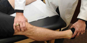
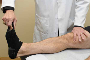
The external rotation recurvatum test is the first test performed to evaluate a patient with potential posterolateral rotatory instability. In this test, the examiner determines if there is an increased amount of knee hyperextension compared to the contralateral side. The test […]
Assessment of Posterolateral Knee Instability
The posterolateral corner of the knee is a very complex area with many important structures which are intricately related to prevent both increased lateral compartment gapping and rotation. There are many different tests which are utilized to assess for posterolateral rotatory instability of the knee. While some examiners feel that the increased amount of varus […]
Anteromedial Drawer Test
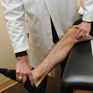
The anteromedial drawer test assesses the amount of increased external rotation due to a medial knee injury. It is performed with the knee flexed to 90°, the foot in external rotation, and an anteromedial rotatory force is applied to the proximal tibia. This test is commonly confused with the posterolateral drawer test (to […]
Medial Knee Instability
The medial, or inside, of the knee is the source of the most frequent knee ligament injury. While commonly referred to as the “MCL”, the medial side of the knee is actually composed of several important structures, with several components, which must be assessed to determine the best way to get back to proper functioning. […]
Tibial Drop Back Sign
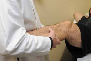
With the tibial drop back sign, one relies on gravity with the knee flexed to 90° to compare if there is any “dropping back” of the tibia on the injured side compared to the normal contralateral knee. In this test, one compares the prominence of the proximal tibia to the femoral condyles with the knee […]

