(Click to Enlarge)
(Click to Enlarge)
(Click to Enlarge)
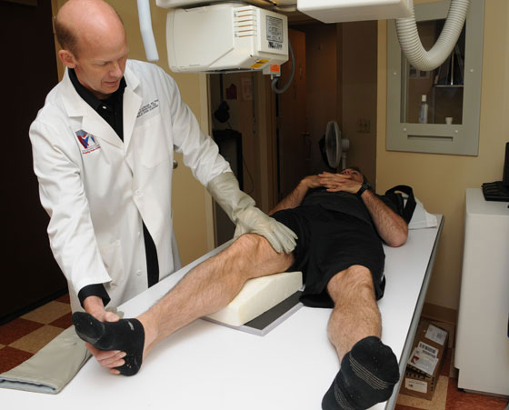
Stress x-rays are an essential component of the workup for a patient with a knee ligament injury. They provide an objective measure for determining the extent of an injury prior to surgery, assessing whether one is healing a partial tear nonoperatively, and in assessing postoperative outcomes. We strongly believe that the use of stress x-rays is necessary to properly diagnose most multiligament knee injuries. This is because it has been clearly demonstrated that one’s fingers cannot always determine the extent of a ligament injury that is present in a patient.
Valgus stress x-rays are performed to determine how much of an injury has occurred to the medial side of the knee. While the MCL is commonly felt to be the main structure on the medial side of the knee, there are actually several structures which interact to provide knee stability. The use of valgus stress x-rays can help determine the extent of injury to these structures and how well one is healing after treatment. We have validated that the amount of gapping present on a side-to-side valgus stress x-ray is 3.2 mm for a complete superficial medial collateral ligament injury and 9.8 mm for all medial knee structures sectioned at 20 degrees of knee flexion. These numbers are very useful to objectively determine the extent of injury and whether one can perform a surgery which has a high success rate of being able to restore stability to the injured knee.
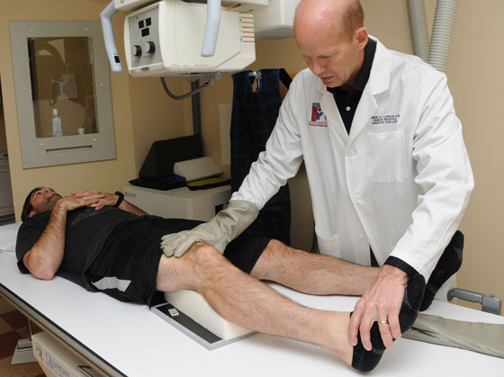
Varus stress x-rays are also essential to determine the extent of a posterolateral knee injury. This is especially important because there can be great variability between patients for the amount of normal lateral compartment gapping that can occur. Thus, these side-to-side stress x-rays, comparing the injured to the non-injured opposite side, are extremely useful to determine if one has a complete posterolateral corner injury.
Our studies have demonstrated that a complete fibular collateral ligament tear demonstrates 2.7 mm of increased gapping, while sectioning of the fibular collateral ligament, popliteus tendon, and popliteofibular ligament, which are the 3 main static stabilizers on the posterolateral aspect of the knee, results in 4.0 mm of increased side-to-side gapping.
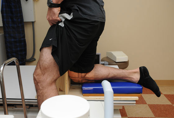
While it is becoming increasingly recognized that valgus and varus stress x-rays are essential to properly diagnose medial-sided and posterolateral knee injuries, it is also becoming increasingly recognized at major multiligament injury centers that the use of PCL stress x-rays are even more important. It has been well documented in clinical care and in the literature that it is very difficult to objectively determine the grade of a PCL injury based on solely the posterior drawer test. Thus, the use of PCL stress x-rays is recommended in all patients with a suspected PCL tear to determine if a patient has a partial PCL tear, a complete isolated PCL tear, or a complete PCL tear with other concurrent ligament injuries. In general, the literature reports that a complete isolated PCL tear has 8 mm to 11 mm of increased posterior translation compared to the normal contralateral side, while a PCL tear with another ligament injury, which usually is either a posterolateral or severe medial-sided knee injury, has 12 mm or more of increased posterior translation compared to the contralateral side. Thus, posterior translation increases of 0 to 6 mm are usually felt to be either due to a partial tear, or may be found in patients with a complete tear who are still too sore after the injury to be able to put full weight on their knee while kneeling and need to be reassessed at a later time to determine the extent of the PCL tear.
There are a few limitations to the use of stress x-rays. First, it is recommended that they performed by someone who has experience in performing the stress x-rays, especially for the ones that require a physician to apply the stress views (for the varus and valgus stress x-rays). This is because an experienced examiner can usually get the patient to relax, even in the face of an acute injury, and a valid objective measure can be determined. The same is true for PCL stress x-rays. If a patient guards, they may not demonstrate the true amount of increased posterior instability. Thus, it is important to discuss with the patient prior to obtaining the PCL stress view that they try to place all their weight on their injured knee’s tibial tubercle for a few seconds while the x-ray is being taken to try to get the best objective measurement possible. The other main issue is that just sometimes patients have so much pain that they cannot relax enough, or they just cannot relax at all even when they do not have pain, to determine the true amount of instability. Thus, what we can definitely say for all patients is that when there is an increased amount of gapping, an injury is present, but when there is no increased gapping seen on the stress x-ray, especially when one thinks on physical exam that there should be, then an assessment must be made as to whether the patient was able to relax enough to be able to physically cooperate with obtaining a stress x-ray view.