Feed Facebook Twitter Linkedin Google Mail

  • Home
  • About
    • Dr. Robert LaPrade
    • The Steadman Clinic
    • Our Team
    • BioMedical Engineering
  • Injuries
    • Articular Cartilage Injuries
    • Complex and Multi-Ligament Injuries
    • Isolated Ligament Injuries
    • Meniscal Injuries
    • Patellar Knee Injuries
    • Soft / Connective Tissue and Nerve Injuries
  • Treatments
    • Arthroscopic Knee Surgery
    • Articular Cartilage Treatment
    • Ligament Reconstructions of the Knee
    • Meniscus Treatment
    • Osteoarthritis Treatment
    • Patellofemoral Joint Treatments
  • Patient Resources
    • Clinical Exam
    • Pre-Operative Info
    • Surgery Information
    • Post-Operative Info
    • Office Information
    • Insurance
    • Patient Forms
    • International Patients
  • Press & Publications
    • Articles & Studies
  • Our Patients
    • Patient Stories
  • News
  • Contact

Inspection

One of the first things a physician does is to inspect the knee. Abrasions, lacerations, previous scars, swelling or prominences are important to evaluate. Swelling around the knee can be either due to causes outside the knee (extraarticular) or causes from inside the knee (intraarticular). In most circumstances, a physical exam can determine whether the swelling is from inside or outside the knee. 

Localized swelling is most often due to an injury. It may indicate a tear of a collateral ligament, bruise or other localized injury. There may also be swelling around the pes anserine bursa, a Baker’s cyst, which is swelling from inside the knee which leaks out through a hole on the back of the knee between the direct arm of the semimembranosus tendon and the medial gastrocnemius tendon, or other causes.
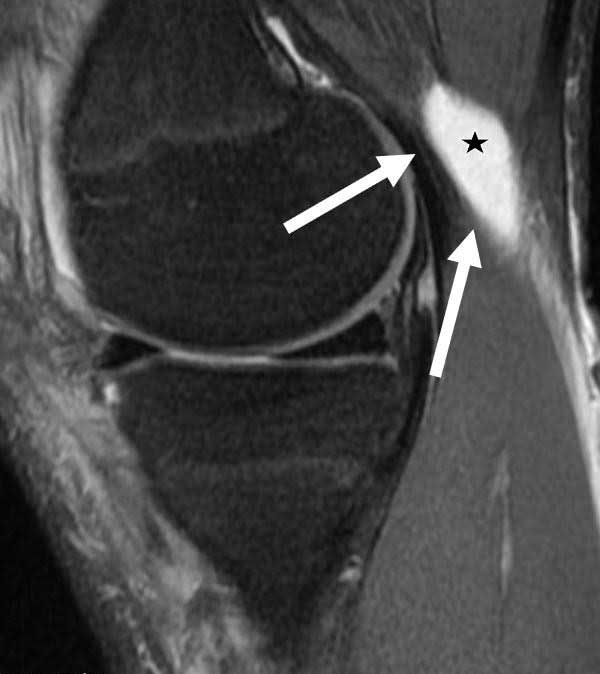
An intraarticular knee effusion can be due to bleeding from an acute injury, gout or pseudogout, swelling from arthritis, infection or other causes. It is important to determine whether the knee is warm, red, or painful to also evaluate intraarticular swelling. Intraarticular knee effusions usually result in the loss of the normal contour of the extensor mechanism, with fullness along the edges of the inside and outside of the quadriceps tendon. Large effusions may also cause the patella to be more mobile. Sometimes, a fluid wave can be created by palpating one side of the joint and feeling the fluid wave hit ones fingers on the other side of the joint. This is not as useful as other means to evaluate for an effusion because these are usually pretty obvious. One of the best ways to evaluate subtle effusions is to have the patient maximally flex their knee and compare one side to the other to see if there is any obvious fluid protrusion along the edges of the patellar tendon.

Tweet
E-mail

For more information about Dr. Robert F. LaPrade, complex orthopedic knee surgeon, please contact his Vail, CO office at: 970.479.5881 or use this contact form:  Contact Dr. Robert LaPrade - Knee Surgeon

Site Search

Clinical Exam

  • History
  • Inspection
  • Palpation of Joint
  • Assessment of Knee ROM
    • ROM – EXT
    • ROM – FLEX
      • Anterior Knee Pain
      • Posterior Knee Pain With Flexion
  • Assessment of Tibiofemoral Joint Line
    • Assessment of Meniscal Tears
  • Assessment of Patellofemoral Joint
    • Patellofemoral Joint Exam
      • Patellar Stability Testing
        • Lateral Patellar Apprehension Test
        • Medial Patellar Apprehension Test
      • Hamstring Tightness Assessment
      • Thigh Circumference Measurement
      • Suprapatella Plica Palpation
  • Assessment of Tibiofemoral Joint
    • Tibiofemoral Joint Exam
      • Anterior Stability Testing
        • Lachmans Test
        • Anterior Drawer Test
        • Pivot Shift Test
        • KT-1000 Test
      • Posterior Stability Testing
        • Tibial Drop Back Sign
        • Quad Active Test
        • Posterior Drawer Test
      • Medial Stability Testing
        • Valgus Stress Test at 0
        • Valgus Stress Test at 30 -Flex
        • Anteromedial Drawer Test
      • Posterolateral Stability Testing
        • External Rotation Recurvatum Test
        • Varus Stress at 0
        • Varus Stress at 30- Flex
        • Posterolateral Drawer Test
        • Dial Test at 30
        • Dial Test at 90
        • Reverse Pivot Shift
        • Figure 4 Test
        • Gait Analysis
  • Objective Use of Stress Radiographs
  • Functional Sports Testing
    • The Sports Test
CONTACT INFO RELATED LINKS DISCLAIMER
Dr. Robert LaPrade

181 West Meadow Drive, Suite 400
Vail, CO 81657

970.479.5881 Dr. LaPrade
970.476.1100  Steadman Clinic
970.479.5835  Fax

Map and Directions
The Steadman Clinic 
Vail Valley Medical
Steadman-Philippon Research Institute
Howard Head Sports Medicine
Kneeguru
AAOS
AOSSM
Oslo Sports Trauma
ESSKA
ISAKOS
All information contained on the drlaprade.com website is intended for informational and educational purposes. The information is not intended nor suited to be a replacement or substitute for professional medical treatment or for professional medical advice relative to a specific medical question or condition. (Website Terms and Conditions and Privacy Policy)

Home | About | Injuries | Treatments | Patient Resources | Press & Publications  | Our Patients | News | Site Map

Copyright © - 2012